
Noninvasive Brain Stimulation Suggests Better Appetite Control in Pilot Study
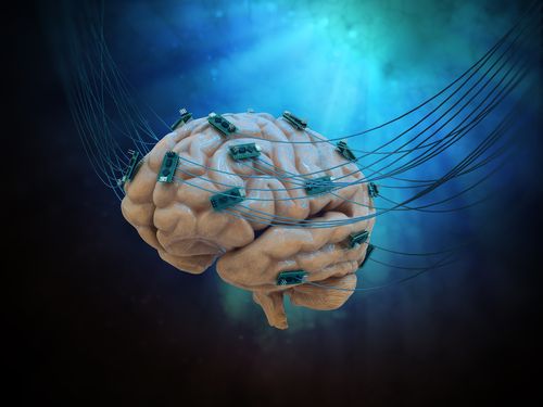


Transcranial direct current stimulation (tDCS) — a non-invasive brain stimulation technique — altered brainwave patterns in people with Prader-Willi syndrome (PWS) in ways consistent with suppressing impulses in response to food, a pilot study in patients reported.
These findings support the use of tDCS to ease the excessive appetite associated with the condition.
The study, “Effects of Transcranial Direct Current Stimulation (tDCS) on Go/NoGo Performance Using Food and Non-Food Stimuli in Patients with Prader–Willi Syndrome,” was published in the journal Brain Sciences.
PWS is caused by the loss of or defects in paternal genes that control intellectual abilities, social behavior, appetite, metabolism, and growth.
Patients develop an excessive appetite (hyperphagia), leading to obesity without sharp caloric restriction programs in place.
tDCS uses painless, weak electrical currents to stimulate specific parts of the brain. It can modulate cognitive and behavioral processes in children and adults.
Studies suggest food decisions are influenced in a brain area called the dorsolateral prefrontal cortex (DLPFC), which has been shown to have low activity in PWS patients when processing responses to food. Targeting this region has been used to lessen cravings for various substances, including tobacco and food, even after a single session.
Researchers at the University of Kansas Medical Center previously showed that tDCS targeting the DLPFC reduced hyperphagia in PWS, as assessed by questionnaires.
They now expanded on this work, testing the impact of DLPFC-directed stimulation on hyperphagia in PWS through performance tasks involving food and non-food images, as well as an electroencephalogram (EEG) to measure brain electrical activity before and after tDCS.
The study included 10 people (six men, four women) with PWS, ranging in age from 19 to 44. Four carried the 15q11-q13 deletion (DEL), in which genes on paternal chromosome 15 are missing, and six had maternal disomy 15 (UPD) — the inheritance of two copies of chromosome 15 from the mother rather than one from each biological parent.
Six participants were randomly assigned to tDCS (2 DEL, 4 UPD), and four (2 DEL, 2 UPD) to a sham tDCS treatment as a control group.
tDCS involved placing two electrodes on the scalp that delivered direct current for 30 minutes. Sham stimulation was delivered with a brief ramp-up and ramp-down of electrical stimulation for 60 seconds at the beginning and end of 30 minutes. EEG was recorded before (baseline) and after tDCS.
Performance tasks started with a food or non-food picture displayed on the computer screen for one second, followed by a black screen. Participants then mouse clicked a button to respond a Go condition for non-food images, and made no clicks to “not respond” to a NoGo condition for food images.
Performance was measured by response times to correct non-food images (Go), the number of correct responses to non-food images (hit rate), and the mistakes — a click response — to food images (NoGo).
At baseline, those assigned to tDCS showed significantly faster reaction times to non-food images compared with the control group. No significant difference between DEL and UPD patients was seen in Go reaction time, hit rate, or incorrect response to NoGo.
Results showed no difference between tDCS treatment or sham groups in Go reaction time. Researchers also found no differences within (comparing after to before intervention) and between these groups in hit rate or incorrect response to NoGo.
EG measures did not lead to differences between the non-food (Go) vs. food (NoGo) images, either before or after intervention in both groups, in a brain wave pattern called N2 amplitude. This pattern has been associated with the ability to suppress responses to stimuli.
No significant differences were found in N2 amplitude in the non-food images between those with a DEL mutation compared with a UPD mutation participants. But the DEL group had significantly smaller N2 amplitudes for the NoGo food condition.
tDCS treatment or sham also did not generate a significant change in N2 amplitude for the Go images. For NoGo food images, however, the tDCS group showed a significant decrease in N2 amplitude after treatment.
Notably, this difference was not observed in the control group, suggesting “tDCS affects response inhibition in this context,” the researchers wrote. A similar reduction in N2 amplitude was associated with a lesser caloric intake after tDCS in women without PWS.
The study “represents a proof of concept that targeted modulation of dysfunctional brain regions associated with attention and response inhibition may be associated with hyperphagia behavior in PWS,” the scientists wrote. “This pattern of results may represent potential neural activity that is associated with subsequent behavior change, which may be of significance” in these patients.
“Given the nature of hyperphagia in this patient population,” added the researchers, the results may reflect changes “in information-processing relating to food stimuli in the human brain that may be associated with additional non-pharmacological intervention opportunities for individuals with PWS.”
Future studies with larger groups of participants are needed to replicate the findings, they said.