
Infants with PWS show early blood flow changes in key brain regions
Findings were seen before later hallmark symptoms fully emerged
Written by |


Infants younger than 6 months with Prader-Willi syndrome (PWS) showed increased blood flow in brain regions involved in movement, motivation and reward, emotional processing, and social behaviors, a study in France reports.
Data also showed that blood flow in one brain region was associated with oral-motor performance, including sucking patterns, and birth weight. Functional connectivity in other brain regions was linked to social functioning and blood levels of unacylated ghrelin, a form of ghrelin involved in metabolism and appetite.

Brain blood flow changes may offer early clues in PWS
“These findings provide a basis for future longitudinal [follow-up] studies to clarify how early patterns of brain function are related to developmental outcomes” in people with PWS, the researchers wrote.
The study, “Early neurodevelopmental brain perfusion abnormalities and functional connectivity findings in infants with Prader-Willi syndrome,” was published in the Journal of Neurodevelopmental Disorders.
PWS is caused by the loss of function of genes located in a region of chromosome 15, called the PWS locus. It is characterized by symptoms such as weak sucking, feeding difficulties, and impaired growth in the first years of life, followed by cognitive and behavioral problems, and hyperphagia, or excessive hunger.
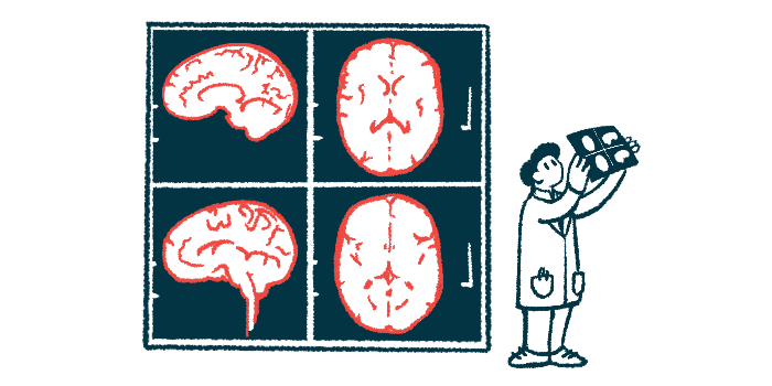
Brain imaging studies in children and adults with PWS have revealed abnormalities in several brain regions, particularly those involved in hunger and appetite regulation. However, little is known about brain development and perfusion, or blood flow in the brain, in infants with the disease.
Here, researchers at the Necker-Enfants Malades Hospital, in Paris, aimed to characterize functional brain organization in infants with PWS younger than 6 months.
MRI study looked at infants younger than 6 months
The study included 15 infants (eight boys and seven girls) with PWS and a mean age of 2.7 months who underwent brain MRI at the researchers’ hospital as part of a clinical trial (NCT04283578). All had early PWS features, including severe low muscle tone, weak sucking, and feeding difficulties, but none had received therapeutic intervention before imaging.
A total of 16 age-matched infants (eight boys and eight girls) who had undergone brain MRI as part of routine care for non-neurological indications were also included.
Brain MRI was conducted during sleep and included arterial spin labeling in both groups to quantify brain blood flow. Resting-state functional MRI was used in the PWS group alone to measure spontaneous brain activity and functional connectivity.
After image quality control, the team analyzed brain blood flow data from 13 infants with PWS and 14 controls, and brain connectivity data from 12 infants with the disease.
Compared with controls, infants with PWS had significantly higher blood flow in five brain clusters that could be grouped into three regions: bilateral insula extending into the superior temporal gyrus (insula-STG), the striatum-pallidum, and the anterior cingulate cortex (ACC).
These regions are involved in movement, motivation and reward, emotional processing, and social behaviors. No brain region in PWS infants showed decreased blood flow relative to controls.
These findings suggest that brain blood flow changes can be detected in the first months of life, before later hallmark PWS features such as excessive hunger are fully evident.
“The insula, ACC, and striatum form a core feeding network with complementary roles: the insula integrates hunger-satiety signals, the ACC regulates motivational control, and the striatum-pallidum drives food-seeking behavior,” the researchers wrote. “Therefore, altered perfusion within these regions may reflect early imbalances in circuits regulating satiety [sensation of being full], impulse control, and reward-driven feeding, potentially contributing to the later development of hyperphagia.”
Blood flow tied to sucking patterns and birth weight
Higher blood flow in the ACC was significantly associated with more dysfunctional oral-motor patterns, shown by lower scores on the Neonatal Oral-Motor Assessment Scale, and with higher birth weight.
Within the PWS group, brain connectivity data showed that greater connectivity between the left and right striatum-pallidum was significantly associated with higher scores on the Coding Interactive Behavior Child State subscale, a measure related to an infant’s mood and interactive state during parent-infant interaction.
This may indicate that functional connectivity “contributes to social regulation during early neurodevelopment,” the researchers wrote.
In addition, higher blood levels of unacylated ghrelin were significantly associated with higher functional connectivity between the insula-STG and the striatum-pallidum.
“This association suggests an early link between metabolic signals and neural circuits involved in feeding and social behavior in individuals with PWS,” the researchers wrote.
Because the insula contributes to pain processing and the ACC is key for emotional regulation, the researchers hypothesized that increased blood flow in these regions could be related to the later emergence of “atypical pain perception” and “poor frustration tolerance and emotional dysregulation frequently observed in this population.”
“These results suggest that perfusion-based measures could support developmental monitoring in PWS and emphasize the importance of longitudinal neuroimaging studies to establish their predictive significance for later outcomes,” the team concluded.


Leave a comment
Fill in the required fields to post. Your email address will not be published.